Personalized Medical Care & Dedicated International Patient Concierge

Medical Guide
Tear Trough Hollowing: Why Under-Eye Aging Looks Dark
Three different things produce the same complaint about dark circles, and only one of them is a shadow. Telling them apart decides what can actually help.
Schedule a ConsultationTear Trough Hollowing: Why Under-Eye Aging Looks Dark
Medically reviewed by Dr. Hong Sung Taek, Chief Director, Merry Plastic Surgery
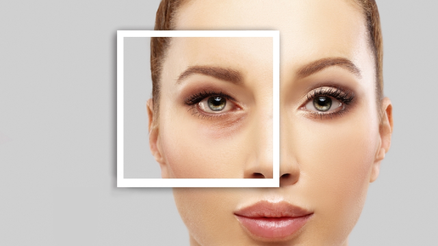
"Dark circles" is the most common complaint about the under-eye area and one of the least useful descriptions in medicine, because at least three unrelated things produce it.
A shadow, cast by a bulge above a groove.
Pigment, in the skin itself.
Transparency, where thin skin lets the darker tissue beneath show through.
They look similar in a bathroom mirror. They respond to completely different things. And the one this article is about — the shadow — is the one most amenable to a structural answer, which is why it dominates surgical discussion and why patients with the other two often end up disappointed by a procedure aimed at it.
What the tear trough actually is
A groove. It runs diagonally from the inner corner of the eye outward and downward, and it exists because the tissue there is tethered — held down against the bone beneath by attachments that do not release.
Above that tether sits orbital fat, cushioning the eye. Below it sits the cheek.
In youth the transition across all three is gradual enough to read as one continuous surface. What changes with time is not usually that the groove deepens on its own, but that the relationship between the three parts alters: the fat above becomes more prominent or shifts forward, the cheek below loses volume and descends, and the tethered line between them becomes the edge of a step rather than a gentle slope.
Which is why the same anatomical feature can be invisible at twenty and defining at forty-five without anything having been added or removed.
The National Library of Medicine's overview of skin aging covers the tissue changes involved, and eyelid disorders the surrounding anatomy.
Why a groove reads as darkness
Because a groove is a place light does not reach.
Overhead light — which is most light, indoors and out — strikes the prominence above and the cheek below, and skips the recess between. The eye interprets the unlit band as pigment rather than as geometry, and reports "dark circles."
The clean test is to change the light. Hold a mirror and tilt your face upward toward a window or a lamp so light comes from below rather than above. If the darkness largely disappears, you were looking at a shadow. If it stays, something in or under the skin is dark, and no amount of contour correction will change that.
That thirty-second test sorts most people into the right conversation, and it is the single most useful thing in this article.
The other two, briefly and honestly
Pigment. Melanin in the skin of the lower lid, which can be constitutional, post-inflammatory, or related to sun exposure. It does not change with the angle of light and it is not a surgical problem. The National Library of Medicine covers skin pigmentation disorders, and sun exposure covers one contributor.
Transparency. The skin of the lower lid is among the thinnest on the body, and it thins further with age. What shows through is the vasculature and tissue beneath, which reads as bluish or purplish rather than brown. Again, not a contour problem.
Most real faces have some of all three. The question at an assessment is not which one you have but which one is doing most of the work, because that determines whether a structural correction will change what bothers you or merely change the shape underneath it.
What the clinic's published assessment weighs
Merry Plastic Surgery's published consultation for this area evaluates the under-eye area to identify the underlying cause, considering:
Fat protrusion. Tear-trough depth. Adhesions. Pigmentation. Hollowing.
Five factors — and notice that pigmentation is one of them, listed alongside the structural ones. That is the assessment doing precisely the sorting this article describes.
And the published output of that assessment is a choice between fat repositioning, fat removal, filler, fat grafting, laser treatment, or another approach.
Six routes. One complaint. The route follows the finding.
Why "adhesions" is on that list
Worth its own paragraph, because it is the least intuitive item and it explains a common disappointment.
The tether that creates the groove is a physical attachment. If it is not released, adding volume above it produces a fuller area that still has a line across it — the groove holds its position because something is holding it.
This is why the clinic's surgical description for this area includes release of tear-trough adhesions as a distinct step before fat is repositioned, and why a correction aimed at the contour has to address the tether rather than only the volume.
If you have previously had something done to this area that made it fuller without making it smoother, that mechanism is a plausible explanation and worth mentioning.
What changes the appearance without changing the anatomy
Three things worth knowing, because they explain day-to-day variation and because two of them are free.
Fluid. The tissue here holds fluid readily, which is why the area looks different on waking, after salt, after alcohol, after crying, and after a long flight. The National Library of Medicine covers edema. A face assessed on the morning after a transpacific flight is not being assessed at baseline — worth knowing if you are flying somewhere for a consultation.
Sleep and fatigue. Which alter both fluid distribution and how you hold your face.
Light, as above. The single biggest source of variation in how bad this looks, and the reason photographs of the same face at the same time can disagree completely.
None of those are reasons to dismiss the concern. They are reasons to judge it under consistent conditions rather than on the worst morning.
What to bring to an assessment
Photographs of yourself in consistent, neutral light, taken straight on and slightly from below. The second angle matters here more than anywhere else on the face, because it changes how a groove reads.
A note of what varies. Worse in the morning? Worse in overhead light? Worse in photographs than in a mirror? Each of those points somewhere.
Your history for this area specifically. Previous filler, previous surgery, previous laser. Filler in particular can persist and change how the area behaves.
An inventory of what you take, read off the packaging. drug safety explains why the list matters; over-the-counter medicines and dietary supplements are the two groups most often left off it by people who do not count them as drugs. Any decision to pause something prescribed belongs to the prescriber.
The honest version of what bothers you. "I look tired in every photograph" and "there is a line under my eye" are different complaints that lead to different assessments.
What this article deliberately does not do
It does not tell you which cause you have. The mirror-and-light test narrows it; an examination decides it.
It does not recommend a treatment. The published assessment lists six possible directions and chooses between them after looking.
It does not promise that any of them will remove darkness. If pigment or transparency is the main contributor, a contour correction changes the contour.
That last point is the one worth carrying into any consultation about this area, anywhere. The most common disappointment in under-eye treatment is not a poor result — it is a good result aimed at the wrong cause.
You can start the conversation before traveling through the clinic's contact page, and the clinic puts its own account of its method on its about page. For the very large amount written about under-eye circles elsewhere, the National Library of Medicine's guidance on evaluating health information and health fraud is worth applying — this is a subject with an unusual density of confident claims.
Why photographs and mirrors disagree
Almost everyone with this complaint has noticed that the problem looks worse in pictures than in the bathroom, or the reverse, and concluded that one of the two is lying. Neither is. They are different instruments.
A mirror gives you a moving, binocular, self-corrected view. You are looking at yourself from a distance you choose, in light you are used to, holding your face the way you hold it when you are looking at yourself — which for most people is subtly more alert than their resting expression.
A photograph freezes one instant under someone else's light. Flash from camera height flattens contour and can erase a groove entirely. Overhead light deepens it. A phone held low changes the geometry of the whole lower lid. And the moment captured is often mid-blink, mid-sentence, or at the end of a long day.
Two practical consequences.
Standardize before you judge. If you want to know what this area actually looks like, take photographs in the same place, at the same time of day, under the same light, from the same two angles — straight on, and slightly from below. Repeat over a week. What survives that is the finding; what does not is variation.
Bring the standardized set, not the worst one. A consultation built around the single most unflattering photograph you own is planning against an outlier. The clinic's published assessment evaluates fat protrusion, tear-trough depth, adhesions, pigmentation and hollowing in person — and consistent photographs support that rather than substituting for it.
One more thing worth knowing: the appearance of this area will not be identical on both sides, and it was not identical before whatever you have noticed began. Asymmetry here is the norm, and an assessment that names it is describing your face accurately rather than finding a new problem.
The Bottom Line
- "Dark circles" describes at least three different things: a shadow cast by a groove, pigment in the skin, and thin skin showing what lies beneath.
- Change the light to sort them. If tilting your face toward a low light source makes the darkness largely disappear, you were looking at geometry rather than color.
- The tear trough is a tethered groove, which is why correcting the contour involves releasing the tether rather than only adding volume above it.
- The clinic's published assessment weighs fat protrusion, tear-trough depth, adhesions, pigmentation and hollowing, and chooses between six possible routes.
- The most common disappointment here is a good result aimed at the wrong cause — so the cause is the thing to establish first.
Frequently Asked Questions
What is the tear trough?
A groove running diagonally from the inner corner of the eye outward, created by tissue tethered down to the bone beneath.
Why does it look dark?
Because a recess does not catch overhead light. The unlit band reads as pigment even though it is geometry.
How can I tell a shadow from pigment?
Change the light. If tilting your face toward a low light source makes the darkness largely disappear, it was a shadow.
Does everyone with dark circles have a tear trough problem?
No. Pigment in the skin and thin skin showing underlying tissue produce similar-looking darkness and are not contour problems.
What does the clinic assess?
Fat protrusion, tear-trough depth, adhesions, pigmentation and hollowing — then chooses between fat repositioning, fat removal, filler, fat grafting, laser treatment or another approach.
Why do adhesions matter?
Because the groove is held down by attachments. Adding volume above an unreleased tether produces fullness with a line still across it.
Why does it look different from day to day?
Fluid, sleep and light. The tissue here holds fluid readily and the appearance changes substantially with the direction of illumination.
What should I bring to a consultation?
Photographs in consistent light from two angles, a note of what makes it worse, your history of filler or surgery in the area, and a written medication list.
This article is for general informational purposes and does not constitute individualized medical advice. It is not a self-diagnosis tool. Surgical results vary with anatomy, tissue condition and individual healing, and whether a procedure is appropriate for you can only be determined through an in-person consultation and examination by a qualified plastic surgeon.
More Articles
Continue reading our clinical guides.

Operating Room Standards: What You Can Ask and Verify
You will never inspect a sterilization room. What you can do is ask questions whose answers reveal whether a facility thinks about this systematically.
Learn More
Staying in Korea After Rhinoplasty: Why No Number Exists
The clinic publishes no fixed figure, and that is not an omission. A nose heals on a timetable long enough that any single number would be misleading.
Learn More

Bringing Someone With You: What a Companion Actually Does
Decide this by listing the hours a companion would actually cover, rather than by how you feel about going alone. Most are ordinary, and a few are not.
Learn More