Personalized Medical Care & Dedicated International Patient Concierge

Medical Guide
The 3D CT Scan Before Rhinoplasty: What Actually Happens
It is the step patients worry about and the one that takes least time. Here is what the machine does, what it looks for, and what it cannot tell anyone.
Schedule a ConsultationThe 3D CT Scan Before Rhinoplasty: What Actually Happens
Medically reviewed by Dr. Hong Sung Taek, Chief Director, Merry Plastic Surgery
Of everything that happens before nasal surgery, the scan is the part patients most often arrive nervous about and the part that is over fastest. It involves no incision, no needle in most planning contexts, and no aftermath.
What makes it worth understanding is not the experience of it. It is what the images are for — because at Merry Plastic Surgery the scan sits at a specific place in a published three-step sequence, and the decisions that follow depend on it.
Step 1 is consultation. Step 2 is diagnostic evaluation. Step 3 is surgical planning. The plan comes after the pictures, not before.
What a CT scan is, in one paragraph
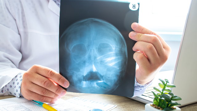
A conventional X-ray sends radiation through you and records what reaches the detector on the other side — one flat image, everything overlapping. A computed tomography scan takes many such measurements from many angles as the equipment rotates, and a computer reconstructs them into cross-sectional slices. Stack the slices and you have a volume, which is what makes a three-dimensional reconstruction possible.
The National Library of Medicine's overviews of CT scans and X-rays cover the underlying method, and MRI scans the alternative that uses magnetic fields rather than radiation and answers different questions.
The practical consequence for a nose: bone, cartilage and airspace can be looked at separately and from any angle, rather than as one superimposed shadow.
Where it sits in the published process
Merry Plastic Surgery's published diagnostic sequence for functional rhinoplasty is explicit:
Step 1 — Consultation. Review of breathing symptoms, discussion of previous trauma or surgery, assessment of cosmetic concerns and goals.
Step 2 — Diagnostic Evaluation. 3D CT imaging, Full HD nasal endoscopy, and evaluation of the septum, turbinates, nasal valve area and sinus anatomy.
Step 3 — Surgical Planning. Identification of the specific structural cause of symptoms, and development of a personalized treatment plan for functional and aesthetic correction when appropriate.
For primary rhinoplasty the published record describes the same tools serving a wider assessment: internal and structural evaluation may include an ENT station, Full HD nasal endoscope, 3D-CT, and Invivo three-dimensional imaging analysis, alongside review of facial proportions, skin thickness, nasal tip position, nostril shape, nasal structure and breathing function, and the relationship of the nose with the forehead, eyes, cheekbones, philtrum, lips and chin.
For revision rhinoplasty, the record adds the reason the scan matters more rather than less: assessment covers inflammation, scar tissue, skin flexibility, implant or foreign material, cartilage support, nasal tip and bridge condition, and internal anatomy, with 3D-CT, an ENT station and Full HD endoscopic equipment used as indicated.
Note the phrasing throughout: "may include," "as indicated." Imaging is part of the assessment toolkit, not an automatic step on every pathway.
Two instruments, two different jobs
Patients often merge the scan and the endoscopy into one idea. They are not the same, and understanding the split makes the appointment legible.
The endoscope looks at surfaces. A camera passed into the nasal passage shows the lining as it actually is — swollen or not, inflamed or not, where the airway narrows in a living, breathing nose.
The scan looks through. It shows structure that no camera can reach: the septum along its full length, the bony architecture of the turbinates, the sinuses behind them, the relationship of everything to everything else.
Neither replaces the other. A septal deviation might be obvious on the scan and invisible at the nostril. Mucosal swelling might dominate the endoscopic view and barely register in the reconstruction. The published record pairs them for that reason, and the surrounding conditions are covered in the National Library of Medicine's overviews of nose injuries and disorders and sinusitis.
What the appointment is like
The published record does not set out a step-by-step account of the scan appointment itself, so what follows describes CT imaging in general terms rather than this clinic's specific protocol. Confirm the details with the team.
Before. You will normally be asked to remove metal from the head and neck area — earrings, glasses, hairpins, piercings, removable dental appliances. Metal in the field scatters the image and can obscure exactly what the scan was ordered to show.
Positioning. For head and sinus imaging you are positioned so that your head is still and aligned. Stillness is the whole requirement. Movement blurs a reconstruction in a way that cannot be corrected afterward, which is why the instruction to stay motionless is repeated.
The scan. The equipment rotates around the region being imaged. You may be asked to hold your breath briefly, or simply to breathe normally and not swallow. It is short — the acquisition is a matter of seconds to a few minutes depending on the equipment and the protocol.
Sensation. There is none, in the ordinary case. No heat, no pressure, no sound beyond the mechanical noise of the equipment. People who find enclosed spaces difficult should say so beforehand; head imaging is generally more open than a full-body scan, but that is a conversation to have in advance rather than while lying down.
After. Nothing. You go about your day. There is no recovery period, no restriction, and nothing to monitor.
The radiation question, answered honestly
CT uses X-rays, which means it uses ionizing radiation. That is a real consideration and it deserves a plain answer rather than reassurance.
Two things are true at once. Imaging is justified when it changes a decision, and this is a case where it does — the published sequence has the scan determining the structural cause that the surgical plan then addresses. And the amount of radiation involved is a legitimate thing to ask about, including whether the scan is necessary in your particular case and whether recent imaging from elsewhere could serve instead.
The National Library of Medicine's overview of radiation exposure covers the general subject. Two practical points follow from it:
Tell the team if you are pregnant or might be. This applies to any imaging using radiation, and it belongs in the conversation before the appointment rather than during it.
Bring prior imaging. The published preparation for revision rhinoplasty asks specifically for previous surgical records, implant information, photographs and imaging when available. An existing scan may answer part of the question and may reduce what needs repeating. Keeping your own copies is worth the effort — personal health records covers how.
What the scan is looking for
Four named structures, from the published record, each of which can independently obstruct breathing:
The septum. The dividing wall between the two nasal passages. Deviation is among the named structural causes of airflow obstruction.
The turbinates. The scrolled shelves on the side wall of each passage. Hypertrophy — enlargement — is another named cause.
The nasal valve area. The narrowest segment of the airway, where narrowing or collapse on inhalation produces obstruction that can be invisible at rest.
The sinuses. The air-filled spaces behind and around the nasal cavity, whose condition affects both symptoms and planning.
Add to that, for aesthetic planning, the bony and cartilaginous framework the published record describes assessing: nasal structure, tip position, cartilage support, and the relationship of the nose to the rest of the face.
One nose. Several possible reasons it does not work. Sorting between them is what the images are for, and it is why the published record says the plan is based on individualized anatomical findings rather than a standardized surgical approach.
What the scan cannot tell anyone
Worth stating, because imaging carries an aura of finality it does not deserve.
It does not decide the operation by itself. It informs a surgeon who is also examining you, listening to your history, and looking at your face as a whole.
It does not show how you will heal. Scar tissue, skin behavior and tissue response are individual, and the published record repeats that healing varies and that outcomes are not promised.
It does not capture the dynamic problem. A valve that collapses on hard inhalation is a moving fault, and a still image of a still nose may not show it. This is why the symptom history — obstruction during exercise or sleep, blockage that changes with position — remains part of the assessment rather than being replaced by pictures.
It does not diagnose inflammation reliably on its own. A nose that is blocked because the lining is swollen from allergy or infection is a different problem from a nose that is blocked by structure, and the published exclusion list names patients with active nasal or sinus infections as needing further assessment before surgery. The National Library of Medicine's overviews of allergy and breathing problems cover that territory.
What to tell the team before you lie down
Six things, and none of them are obvious enough to leave unsaid.
Whether you are or might be pregnant.
Any previous nasal surgery, including how long ago and what was used. The published preparation asks for exactly this.
Any nasal trauma, even old and apparently minor. A break in childhood is relevant anatomy.
Any implant or foreign material placed at any time, anywhere in the nose.
Chronic rhinitis or sinus problems, which the published pre-procedure preparation names directly.
Claustrophobia, difficulty lying still, or anything that makes staying motionless hard — a tremor, back pain, a cough. Better addressed by planning than by endurance.
The clinic's hours are opening is 9:30 a.m. throughout; closing is 6:30 p.m. on weekdays and 4:00 p.m. on Saturdays; Sundays and public holidays are closed, which is worth knowing when scheduling imaging and consultation into a short trip. You can raise the list in advance through the clinic's contact page, and the clinic's own framing of how it works lives on its about page. The National Library of Medicine's guidance on talking with your doctor covers how to make a short appointment carry a lot of information.
The Bottom Line
- The scan sits at Step 2 of a published three-step sequence — consultation, diagnostic evaluation, then surgical planning. The plan follows the images.
- A CT builds cross-sections from many angles, which is what allows bone, cartilage and airspace to be examined separately rather than as one overlapping shadow.
- The endoscope and the scan do different jobs: one shows the living surface, the other shows structure no camera can reach. The published record uses both.
- The appointment itself is short, sensationless and has no recovery; the real preparation is removing metal, staying still, and disclosing history.
- It uses ionizing radiation, so pregnancy and prior imaging are both worth raising — and the scan still cannot show how you will heal or capture a fault that only appears in motion.
Frequently Asked Questions
What does a 3D CT scan show that an examination cannot?
Structure behind and beneath the surface — the septum along its length, the bony turbinates, the nasal valve area and the sinuses — viewable separately and from any angle.
Why do I need the endoscopy as well?
Because the two answer different questions. The endoscope shows the living lining and where the airway narrows in a breathing nose; the scan shows underlying structure.
Does it hurt?
No. In the ordinary planning context there is no incision, no injection and no sensation beyond lying still while the equipment rotates.
How long does it take?
The acquisition is short — seconds to a few minutes depending on equipment and protocol. The appointment around it is longer than the scan itself.
What should I remove beforehand?
Metal in the head and neck area: earrings, glasses, hairpins, piercings and removable dental appliances, since metal scatters the image.
Is the radiation a concern?
CT uses ionizing radiation, so it is a fair question to ask — including whether the scan is needed in your case and whether existing imaging could serve. Tell the team if you are or might be pregnant.
Can I bring a scan done at home?
Yes, and you should. The published preparation asks for previous surgical records, implant information, photographs and imaging when available.
Does the scan decide my surgery?
No. It identifies the structural cause, which the surgeon then combines with examination, symptom history and facial assessment to build an individualized plan.
This article is for general informational purposes and does not constitute individualized medical advice. It is not a self-diagnosis tool. Surgical results vary with anatomy, tissue condition and individual healing, and whether a procedure is appropriate for you can only be determined through an in-person consultation and examination by a qualified plastic surgeon.
More Articles
Continue reading our clinical guides.
Operating Room Standards: What You Can Ask and Verify
You will never inspect a sterilization room. What you can do is ask questions whose answers reveal whether a facility thinks about this systematically.
Learn More

Staying in Korea After Rhinoplasty: Why No Number Exists
The clinic publishes no fixed figure, and that is not an omission. A nose heals on a timetable long enough that any single number would be misleading.
Learn More

Bringing Someone With You: What a Companion Actually Does
Decide this by listing the hours a companion would actually cover, rather than by how you feel about going alone. Most are ordinary, and a few are not.
Learn More